Scientists Create Artificial Embryonic Node to Solve Mystery of Heart and Liver Placement
Eindhoven, Friday, 3 April 2026.
Dutch researchers have built a microscopic artificial embryonic node that reveals why human organs develop on specific sides of the body. Using magnetic artificial cilia just 23 micrometers long, the team recreated the tiny fluid-filled cavity where organ positioning begins during early embryonic development. Their groundbreaking work shows that rotating hair-like structures create precise fluid flows that direct whether organs like the heart grow on the left or liver on the right, solving a fundamental puzzle of human development through innovative bioengineering.
Breakthrough in Developmental Biology Research
This research represents a significant advancement in developmental biology and biomedical engineering. The study, published in Science Advances on March 25, 2026, emerged from a collaborative effort between Eindhoven University of Technology (TU/e) and the University of Groningen [1][2][3]. The research team, led by Professor Jaap den Toonder from TU/e’s Department of Mechanical Engineering and chair of the Microsystems section, successfully created an artificial version of the embryonic node to study how organ asymmetry develops in vertebrates [1][4]. The project represents the culmination of den Toonder’s first ERC Advanced Grant-funded research, which he received in 2019 [1].
Understanding the Embryonic Node Mechanism
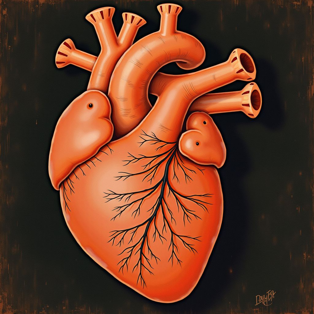
The embryonic node is a critical structure in early vertebrate development, appearing as a small fluid-filled cavity measuring only a few hundred micrometers across [1][2]. In mouse embryos, this structure develops as a closed triangular cavity on the ventral side, measuring 10 to 20 micrometers deep and 50 to 100 micrometers across [6]. Within this tiny space, hundreds of microscopic hair-like structures called cilia perform a synchronized dance that determines the fundamental architecture of the developing body [1][2]. As Professor den Toonder explains, “The cilia in the embryonic node rotate in the same direction, making a tilted conical motion. This generates an anticlockwise fluid flow inside the node, and it’s this flow that is known to play a key role in the left-right symmetry” [1][2][5]. These natural cilia have an average length and diameter of 5 and 0.3 micrometers respectively, rotating at approximately 10 Hz to produce the crucial nodal flow [6].
Engineering an Artificial Solution
The TU/e team overcame the challenge of studying this microscopic process by creating an artificial embryonic node using advanced microfabrication techniques [1][2]. The artificial cilia were crafted from a unique magnetic polymer material developed in TU/e’s Microfab/lab, with each cilium measuring 1 micrometer in radius and 23 micrometers in length [1][3]. These synthetic structures contain 38 ± 2 percent by weight of magnetic particles, allowing researchers to control their movement using magnetic fields [6]. Postdoctoral researcher Tanveer ul Islam, who led the fabrication process, utilized polycarbonate track-etched micro/nanoporous membranes as sacrificial molds to create these precision structures [2][6]. The artificial nodes themselves were made approximately 500 micrometers across - 5 times larger than real embryonic nodes to enable detailed visualization and study [3][5].
Computational Modeling Reveals Hidden Mechanisms
The experimental work at TU/e was complemented by sophisticated computational modeling at the University of Groningen, led by researchers Ishu Aggarwal and Patrick Onck [1][2][5]. The Groningen team developed specialized algorithms to simulate the complex fluid dynamics within the embryonic node, a computational challenge that required months of processing time on high-performance supercomputing facilities [2][5]. Professor Onck, who has collaborated with den Toonder for over 20 years on magnetic artificial cilia research, noted the complexity of the simulation: “Many algorithms discretize the fluid into a mesh with very small elements, but in three dimensions it gets harder to solve the system - especially for an embryonic node at the micrometer scale that consists of over hundreds of cilia” [2][5]. The simulations revealed that the artificial node produces dissimilar fluid velocity profiles around primary cilia on the left and right sides, creating distinct cilium bending patterns and asymmetric distribution of signaling particles [6].
Implications for Medical Understanding and Future Applications
The research demonstrates that two synergistic mechanisms work together to break left-right symmetry during embryonic development: mechanical sensing through cilia bending and chemical sensing through morphogen distribution [2][5][6]. This finding represents what den Toonder calls “an eyeopener for the developmental biologists” with significant impact on the scientific understanding of embryonic development [4]. While the research is fundamentally curiosity-driven, it opens new avenues for understanding congenital conditions related to organ positioning [4]. Den Toonder acknowledges that direct intervention in embryonic development would be complex, but the artificial embryonic node is now available for biologists, clinicians, and other researchers to explore further applications [1][4]. Moving forward, den Toonder is focusing on his second ERC Advanced Grant project, which began in early 2026, to study how cancer cells use blood vessels to spread throughout the body [1][4].