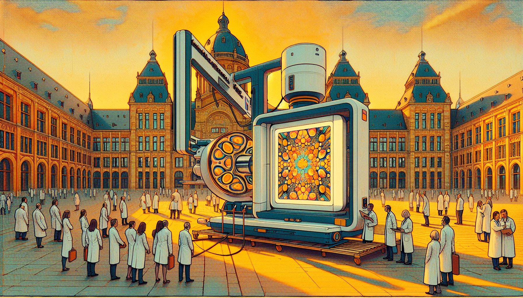
TU/e Develops Game-Changing Compact X-ray Machine for Art and Science

Eindhoven, Friday, 21 March 2025.
Researchers at Eindhoven University have unveiled a compact X-ray machine, revolutionizing painting analysis with adjustable X-rays, enabling studies of layered artworks and offering potential in medical imaging.
Breakthrough in Compact X-ray Technology
In a significant scientific advancement, researchers at Eindhoven University of Technology (TU/e) have successfully developed a working prototype of a compact hard X-ray machine. The breakthrough came under the leadership of Professor Jom Luiten and his team, including PhD students Ids van Elk and Coen Sweers, who achieved this milestone in September 2024 [1]. The prototype, housed in the basement of the Qubit building, measures just 1.5 by 3 meters, representing a remarkable achievement in miniaturization compared to traditional X-ray facilities [1]. This innovation is particularly noteworthy as X-rays, discovered by Wilhelm Conrad Röntgen in 1895, have historically required large-scale facilities for high-quality imaging [2].
Revolutionary Applications in Art Analysis
The machine’s most distinctive feature is its ability to generate precise, adjustable hard X-rays, making it particularly valuable for analyzing layered paintings and archaeological artifacts [1]. This capability has significant implications for art historians and conservators, who previously relied on massive facilities like the European Synchrotron Radiation Facility (ESRF) in Grenoble, France, for detailed art analysis [1]. The compact nature of this device means museums and research institutions could potentially conduct sophisticated X-ray analysis on-site, dramatically improving accessibility to this crucial research tool.
Technical Innovation and Safety Features
The development process, which began in 2018 with funding from Interreg Flanders-Netherlands, incorporated essential safety features including lead bulkheads and concrete walls for radiation shielding [1]. This attention to safety is crucial given that X-rays can cause DNA damage and other health hazards [2]. The machine’s innovative design allows researchers to adjust wavelengths with a simple rotary knob, enabling precise matching to specific materials or objects under study [1].
Future Prospects and Collaborative Development
The project’s next phase involves collaboration with partners from TU Delft, University of Antwerp, and Ghent University, focusing on radiation targeting, advanced detection equipment, and data analysis [1]. Beyond art analysis, the technology shows promise in various applications, including silicon wafer inspection, atherosclerosis research, and examination of lung tissue with early-stage COVID damage [1]. The project exemplifies successful international scientific collaboration, supported by Interreg Flanders-Netherlands, demonstrating the potential for breakthrough innovations in X-ray technology [1].

