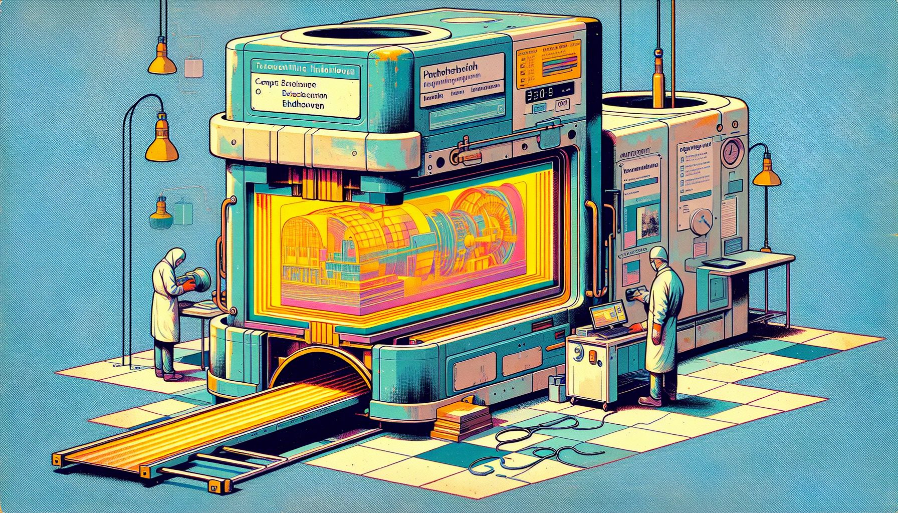
Eindhoven Scientists Develop Compact X-ray Machine for Art Analysis

Eindhoven, Thursday, 19 December 2024.
TU/e researchers have created a compact X-ray machine, enhancing art analysis by revealing hidden layers in paintings, offering an alternative to large, costly synchrotrons.
Breakthrough in Compact X-ray Technology
Scientists at Eindhoven University of Technology (TU/e) achieved a significant breakthrough in September 2024 when PhD students Ids van Elk and Coen Sweers successfully operated their compact hard X-ray machine [1]. Led by Professor Jom Luiten and Peter Mutsaers, the team developed a device measuring just 1.5 by 3 meters [1], drastically smaller than traditional synchrotron facilities like the ESRF in Grenoble [1]. This innovation represents a major advancement in X-ray imaging technology, which has been a crucial tool for non-invasive imaging since its discovery in the 1890s [2].
Revolutionary Applications in Art Analysis
The compact X-ray machine’s primary application lies in high-resolution analysis of art and archaeological objects [1]. Working in collaboration with art historian and materials scientist Joris Dik, known for discovering hidden works by Van Gogh, Rembrandt, and Magritte through X-ray analysis, the team has developed technology that enables detailed study of paint layers and hidden artwork [1]. This advancement is particularly significant as current access to such technology has been limited to large synchrotron facilities, which are typically expensive and fully booked [1].
Technical Innovation and Safety
The prototype, housed in TU/e’s Qubit lab, represents a significant achievement in particle accelerator miniaturization [1]. The facility incorporates comprehensive safety measures, including lead bulkheads and concrete walls to ensure proper radiation containment [1]. The device produces adjustable hard X-rays, offering unprecedented flexibility in imaging applications [1]. This development builds upon decades of X-ray imaging evolution, which has progressed from film-screen radiography to advanced digital systems [2].
Future Implications and Development
The project, initiated in 2018 with funding from Interreg Flanders-Netherlands and government contributions [1], has established collaborations with TU Delft and universities in Antwerp and Ghent [1]. Beyond art analysis, the technology shows promise for various applications, including silicon wafer examination, atherosclerosis research, and lung tissue analysis [1]. The team is currently focused on increasing intensity and improving beam focus, with immediate plans to create a proof of concept for examining paintings [1]. As research partner Hessel Castricum notes, ‘As far as the application is concerned, it is only now that things are starting’ [1].

