
Dutch Innovation Improves Lung Cancer Detection Without Invasive Surgery
Eindhoven, Thursday, 5 November 2026.
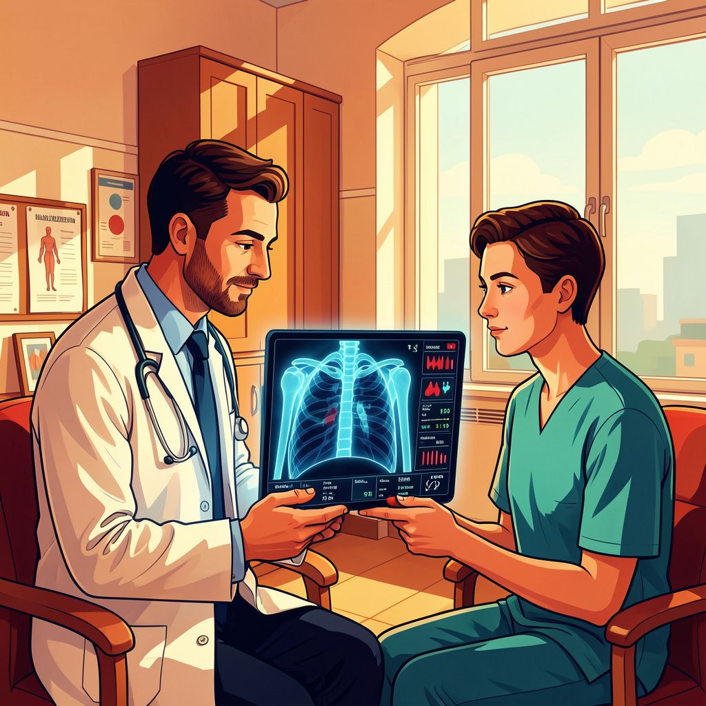
Pairing routine blood tests with chest scans boosted lung cancer detection rates from 62% to roughly 78%, presenting a safer, faster diagnostic alternative for high-risk patients.
Transforming Diagnostics Through Healthtech Synergy
This advancement falls squarely in the healthtech and medical technology domain, addressing one of the most critical bottlenecks in clinical oncology: the reliance on invasive tissue extraction [1][2]. Traditional lung cancer diagnoses often require surgical or needle biopsies, procedures that present physiological risks, induce patient distress, and frequently face delays when a patient is frail or a tumor is anatomically inaccessible [2][7]. By synthesizing non-invasive liquid biomarkers with imaging data inside an analytical computer model, the innovation provides an accurate diagnostic pathway that minimizes procedural trauma and shortens clinical pathways [1][2][5].
Clinical Mechanism and Diagnostic Performance
The technology works by integrating two independent streams of diagnostic insight: serum tumor markers evaluated via standard blood draws and radiographic patterns captured through computed tomography (CT) scans [1][2][7]. In a clinical study analyzing 346 patients separated into two cohorts, evaluating blood tests in isolation yielded a cancer detection accuracy of 62 percent [1][5]. However, when clinical data from blood work were paired with a radiologist’s assessment of CT scans, detection rates rose to approximately 78 percent—representing an absolute accuracy gain of 16 percentage points and a relative diagnostic improvement of 25.806 percent [1][5].
The Dual Role of Artificial Intelligence and Human Expertise
To assess how automation compares to specialist evaluation, researchers implemented a deep-learning artificial intelligence model capable of processing CT scan patterns autonomously [2][5]. When this AI framework was merged with blood test data, it identified lung cancer with 76 percent accuracy in the initial patient group and 73 percent in a second independent validation group [5]. While this demonstrates that algorithmic modeling can achieve results close to the 78 percent achieved by clinical radiologist evaluations, the team emphasized that human expertise remains invaluable in complex diagnostic environments [1][4][5].
Eindhoven Leadership and Clinical Roadmap
The project was spearheaded in Eindhoven, the Netherlands, through a collaboration between Catharina Hospital (Catharina Ziekenhuis) and Eindhoven University of Technology (TU/e) [1][2][5]. The research effort is led by 29-year-old PhD candidate Hao Cao from the General Clinical Laboratory at Catharina Hospital, working alongside TU/e associate professor Fons van der Sommen, PhD candidate Cris Claessens, biomedical engineer Jasper Stiekema, and clinical specialists including Volkher Scharnhorst, Federica Eduati, Christi Steendam, Ben van den Borne, Rianne Verhees, and Astrid Donkers [1][2][5]. In late October 2026, Cao presented these outcomes at the European Society for Medical Oncology (ESMO) Congress in Madrid, where he received an ESMO Merit Award [1][5].
Translational Horizons and Ongoing Research
While the multidisciplinary team stresses that the multi-modal tool does not yet entirely eliminate conventional biopsies, ongoing clinical validation aims to bridge the methodology into daily hospital workflows [3][4]. Cao had previously earned distinction at Catharina Hospital’s Science Evening for research accelerating cancer identification in pleural fluid and secured a €45,000 grant from the Catharina Health Foundation to analyze tumor DNA abnormalities in blood [1][5][7]. Research under this €45,000 foundation project is scheduled for publication later in 2026, advancing precision medicine for vulnerable cancer patients [1][2][5].
Bronnen
- www.tue.nl
- www.tue.nl
- nl.linkedin.com
- www.linkedin.com
- www.myscience.org
- www.facebook.com
- www.catharinaziekenhuis.nl
- www.facebook.com